| Basilar artery | |
|---|---|
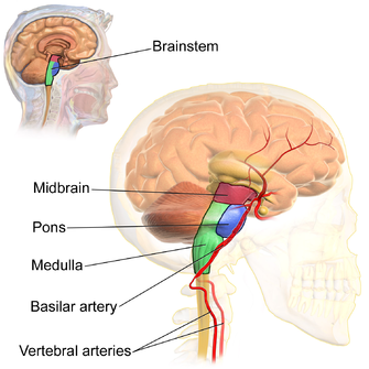
 The basilar artery lies at the front of the brainstem in the midline and is formed from the union of the two vertebral arteries. | |
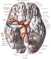
 Diagram of the arterial circulation at the base of the brain (inferior view). The basilar artery terminates by splitting into the left and right posterior cerebral arteries. | |
| Details | |
| Source | Vertebral arteries |
| Branches | Pontine arteries anterior inferior cerebellar (AICA) Paramedian arteries superior cerebellar arteries terminal posterior cerebral arteries. |
| Supplies | Pons, and superior and inferior aspects of the cerebellum. |
| Identifiers | |
| Latin | arteria basilaris |
| MeSH | D001488 |
| TA98 | A12.2.07.081 |
| TA2 | 4548 |
| FMA | 50542 |
| Anatomical terminology | |
The basilar artery (U.K.: /ˈbæz.ɪ.lə/;[1][2] U.S.: /ˈbæs.ə.lər/[3]) is one of the arteries that supplies the brain with oxygen-rich blood.
The two vertebral arteries and the basilar artery are known as the vertebral basilar system, which supplies blood to the posterior part of the circle of Willis and joins with blood supplied to the anterior part of the circle of Willis from the internal carotid arteries.[4][5][6]


