No higher resolution available.
Fluorescent_labeling_of_Bicoid_GFP_and_mRNA.pdf (322 × 466 pixels, file size: 71 KB, MIME type: application/pdf)
| This is a file from the Wikimedia Commons. Information from its description page there is shown below. Commons is a freely licensed media file repository. You can help. |
Summary
| Description |
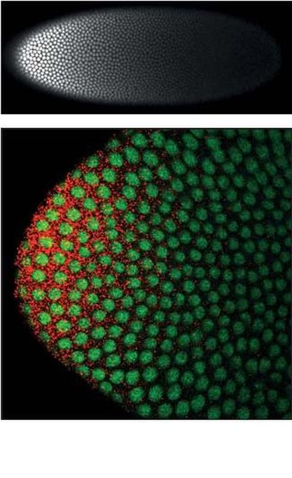
English: Bicoid mRNA is localized to the anterior pole of the Drosophila oocyte (St. Johnston et al., 1989). Bicoid protein enters the nuclei and forms a nuclear gradient from anterior to posterior (Driever and Nusslein-Volhard, 1988). The top photograph shows the nuclear Bicoid protein gradient in a fixed transgenic Drosophila embryo carrying a bicoid–GFP fusion gene. In these embryos the concentration of Bicoid protein could be accurately measured either by the intensity of the autofluorescence of the GFP protein itself (not shown) or by the intensity of fluorescence of an anti-GFP antibody tagged with a fluorescent dye, as shown in a surface view of the embryo here. The lower photograph shows the anterior tip of a fixed transgenic Drosophila embryo carrying a bicoid–GFP fusion gene labeled with fluorescently-tagged cDNA probes that bind bicoid mRNA molecules. Green color shows autofluorescence of the GFP protein in the nuclei, red color shows packages of multiple bicoid mRNA molecules in the anterior cytoplasm. Methods described in Little et al. (2011).
References: Driever, W.; Nüsslein-Volhard, C. (1988). "The bicoid protein determines position in the Drosophila embryo in a concentration-dependent manner". Cell 54: 95. doi:10.1016/0092-8674(88)90183-3 St Johnston D, Driever W, Berleth T, Richstein S, Nusslein-Volhard C (1989). Multiple steps in the localization of bicoid RNA to the anterior pole of the Drosophila oocyte. Development 107 Suppl: 13–19. Little SC, Tkačik G, Kneeland T, Wieschaus EF, Gregor T (2011). The formation of the Bicoid morphogen gradient requires protein movement from anteriorly localized mRNA. PLoS Biology 9(3), e1000596. |
| Source | Sent via email by Thomas Gregor (Principal Investigator) |
| Author | Thomas Gregor, Julien O. Dubuis, Shawn C. Little |
| Permission (Reusing this file) |
This file is licensed under the Creative Commons Attribution-Share Alike 3.0 Unported license.
Attribution: Thomas Gregor, Julien O. Dubuis, Shawn C. Little
- You are free:
- to share – to copy, distribute and transmit the work
- to remix – to adapt the work
- Under the following conditions:
- attribution – You must give appropriate credit, provide a link to the license, and indicate if changes were made. You may do so in any reasonable manner, but not in any way that suggests the licensor endorses you or your use.
- share alike – If you remix, transform, or build upon the material, you must distribute your contributions under the same or compatible license as the original.
Licensing
| This file is made available under the Creative Commons CC0 1.0 Universal Public Domain Dedication. | |
| The person who associated a work with this deed has dedicated the work to the public domain by waiving all of their rights to the work worldwide under copyright law, including all related and neighboring rights, to the extent allowed by law. You can copy, modify, distribute and perform the work, even for commercial purposes, all without asking permission.
|
