| External vertebral venous plexuses | |
|---|---|
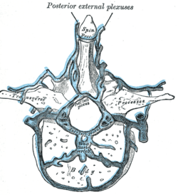
 Transverse section of a thoracic vertebra, showing the vertebral venous plexuses. | |
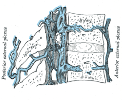
 Median sagittal section of two thoracic vertebrae, showing the vertebral venous plexuses. | |
| Details | |
| Identifiers | |
| Latin | plexus venosi vertebrales externi |
| TA98 | A12.3.07.019 A12.3.07.020 |
| TA2 | 4951, 4952 |
| FMA | 12851 |
| Anatomical terminology | |
The external vertebral venous plexuses (extraspinal veins) consist of anterior and posterior plexuses which anastomose freely with each other. They are most prominent in the cervical region[1] where they form anastomoses with the vertebral, occipital, and deep cervical veins.[2]