In biology
In biological systems, methylation is accomplished by enzymes. Methylation can modify heavy metals and can regulate gene expression, RNA processing, and protein function. It is a key process underlying epigenetics. Sources of methyl groups include S-methylmethionine, methyl folate, methyl B12.[1]
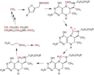
Methanogenesis
Methanogenesis, the process that generates methane from CO2, involves a series of methylation reactions. These reactions are caused by a set of enzymes harbored by a family of anaerobic microbes.[2]

In reverse methanogenesis, methane is the methylating agent.[3]
O-methyltransferases
|
Main article: O-methyltransferase |
A wide variety of phenols undergo O-methylation to give anisole derivatives. This process, catalyzed by such enzymes as caffeoyl-CoA O-methyltransferase, is a key reaction in the biosynthesis of lignols, percursors to lignin, a major structural component of plants.
Plants produce flavonoids and isoflavones with methylations on hydroxyl groups, i.e. methoxy bonds. This 5-O-methylation affects the flavonoid's water solubility. Examples are 5-O-methylgenistein, 5-O-methylmyricetin, and 5-O-methylquercetin (azaleatin).
Proteins
Along with ubiquitination and phosphorylation, methylation is a major biochemical process for modifying protein function. The most prevalent protein methylations affect arginine and lysine residue of specific histones. Otherwise histidine, glutamate, asparagine, cysteine are susceptible to methylation. Some of these products include S-methylcysteine, two isomers of N-methylhistidine, and two isomers of N-methylarginine.[4]
Methionine synthase

Methionine synthase regenerates methionine (Met) from homocysteine (Hcy). The overall reaction transforms 5-methyltetrahydrofolate (N5-MeTHF) into tetrahydrofolate (THF) while transferring a methyl group to Hcy to form Met. Methionine Syntheses can be cobalamin-dependent and cobalamin-independent: Plants have both, animals depend on the methylcobalamin-dependent form.
In methylcobalamin-dependent forms of the enzyme, the reaction proceeds by two steps in a ping-pong reaction. The enzyme is initially primed into a reactive state by the transfer of a methyl group from N5-MeTHF to Co(I) in enzyme-bound cobalamin ((Cob), also known as vitamine B12)) , , forming methyl-cobalamin(Me-Cob) that now contains Me-Co(III) and activating the enzyme. Then, a Hcy that has coordinated to an enzyme-bound zinc to form a reactive thiolate reacts with the Me-Cob. The activated methyl group is transferred from Me-Cob to the Hcy thiolate, which regenerates Co(I) in Cob, and Met is released from the enzyme.[5]
Heavy metals: arsenic, mercury, cadmium
Biomethylation is the pathway for converting some heavy elements into more mobile or more lethal derivatives that can enter the food chain. The biomethylation of arsenic compounds starts with the formation of methanearsonates. Thus, trivalent inorganic arsenic compounds are methylated to give methanearsonate. S-adenosylmethionine is the methyl donor. The methanearsonates are the precursors to dimethylarsonates, again by the cycle of reduction (to methylarsonous acid) followed by a second methylation.[6] Related pathways are found in the microbial methylation of mercury to methylmercury.
Epigenetic methylation
DNA methylation
DNA methylation is the conversion of the cytosine to 5-methylcytosine. The formation of Me-CpG is catalyzed by the enzyme DNA methyltransferase. In vertebrates, DNA methylation typically occurs at CpG sites (cytosine-phosphate-guanine sites—that is, sites where a cytosine is directly followed by a guanine in the DNA sequence). In mammals, DNA methylation is common in body cells,[7] and methylation of CpG sites seems to be the default.[8][9] Human DNA has about 80–90% of CpG sites methylated, but there are certain areas, known as CpG islands, that are CG-rich (high cytosine and guanine content, made up of about 65% CG residues), wherein none is methylated. These are associated with the promoters of 56% of mammalian genes, including all ubiquitously expressed genes. One to two percent of the human genome are CpG clusters, and there is an inverse relationship between CpG methylation and transcriptional activity. Methylation contributing to epigenetic inheritance can occur through either DNA methylation or protein methylation. Improper methylations of human genes can lead to disease development,[10][11] including cancer.[12][13]
In honey bees, DNA methylation is associated with alternative splicing and gene regulation based on functional genomic research published in 2013.[14] In addition, DNA methylation is associated with expression changes in immune genes when honey bees were under lethal viral infection.[15] Several review papers have been published on the topics of DNA methylation in social insects.[16][17]
RNA methylation
RNA methylation occurs in different RNA species viz. tRNA, rRNA, mRNA, tmRNA, snRNA, snoRNA, miRNA, and viral RNA. Different catalytic strategies are employed for RNA methylation by a variety of RNA-methyltransferases. RNA methylation is thought to have existed before DNA methylation in the early forms of life evolving on earth.[18]
N6-methyladenosine (m6A) is the most common and abundant methylation modification in RNA molecules (mRNA) present in eukaryotes. 5-methylcytosine (5-mC) also commonly occurs in various RNA molecules. Recent data strongly suggest that m6A and 5-mC RNA methylation affects the regulation of various biological processes such as RNA stability and mRNA translation,[19] and that abnormal RNA methylation contributes to etiology of human diseases.[20]
In social insects such as honey bees, RNA methylation is studied as a possible epigenetic mechanism underlying aggression via reciprocal crosses.[21]
Protein methylation
Protein methylation typically takes place on arginine or lysine amino acid residues in the protein sequence.[22] Arginine can be methylated once (monomethylated arginine) or twice, with either both methyl groups on one terminal nitrogen (asymmetric dimethylarginine) or one on both nitrogens (symmetric dimethylarginine), by protein arginine methyltransferases (PRMTs). Lysine can be methylated once, twice, or three times by lysine methyltransferases. Protein methylation has been most studied in the histones. The transfer of methyl groups from S-adenosyl methionine to histones is catalyzed by enzymes known as histone methyltransferases. Histones that are methylated on certain residues can act epigenetically to repress or activate gene expression.[23][24] Protein methylation is one type of post-translational modification.
Evolution
Methyl metabolism is very ancient and can be found in all organisms on earth, from bacteria to humans, indicating the importance of methyl metabolism for physiology.[25] Indeed, pharmacological inhibition of global methylation in species ranging from human, mouse, fish, fly, roundworm, plant, algae, and cyanobacteria causes the same effects on their biological rhythms, demonstrating conserved physiological roles of methylation during evolution.[26]


